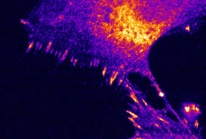
Zebrafish is a valuable model for studying the vertebrate visual system. The transparency of zebrafish during its early development allows the use of fluorescent microscopy. We investigated the optic tectum’s functional development by imaging neural activity from three to five days post-fertilization (dpf), when the zebrafish visual system rapidly develops. We performed fast functional live cell imaging of neural activity using a genetically encoded calcium indicator expressed in the central nervous system of zebrafish larvae. Comparing the neural response to visual stimuli between fish exposed to and deprived of visual information during development, we evaluated the function impact of visual experience in the optic tectum.
